
A CT or CAT scan is a type of X-ray imaging test that takes detailed cross-sectional pictures of the inside of the body. CT scans have become an important diagnostic tool for detecting many types of cancers. The scans allow doctors to see inside the body in great detail, revealing abnormalities that may indicate cancer. CT scans can help detect many cancers at early stages when they are most treatable.
While CT scans are helpful for cancer detection, they do have some limitations. CT scans alone cannot definitively diagnose cancer. They can only detect abnormal structures that may require further testing. Factors like the size of a tumor and skills of the radiologist can also impact the accuracy of CT scans for detecting cancer. Therefore, CT scans are often used along with other tests like biopsies or PET scans to confirm a cancer diagnosis.
What is a CT or CAT Scan?
A CT or CAT scan is a type of medical imaging procedure that uses X-rays and computers to create detailed cross-sectional images of the inside of the body (Cleveland Clinic, 2023). CT stands for “computed tomography.” It is also sometimes referred to as CAT scan, which stands for “computerized axial tomography.”
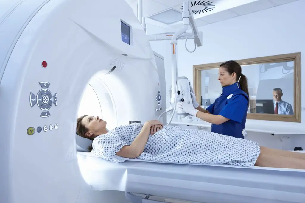
During a CT scan, the patient lies on a table that slides into a large X-ray machine called a CT scanner. The scanner rotates around the body and takes multiple X-ray images from different angles. A computer then processes these images and combines them to produce cross-sectional views of the internal organs and tissues (Hopkins Medicine).
Because it provides more detailed images than regular X-rays, a CT scan allows doctors to see inside the body’s structures including the bones, blood vessels, and soft tissues. This helps diagnose medical conditions and guide treatment.
Uses of CT Scans
CT scans have a wide range of uses in the medical field. Some of the most common uses include diagnosing diseases, guiding biopsies, monitoring cancer treatment, detecting tumors, evaluating organs for donation, diagnosing kidney stones, detecting bone fractures that may not show up on x-rays, showing internal injuries and bleeding, evaluating blood vessels and arteries, finding heart disease and evaluating the extent of plaque buildup, checking for pulmonary embolisms, detecting aneurysms, pinpointing areas of infection, and planning radiation therapy for cancer treatment.
CT scans provide detailed cross-sectional images of the body, allowing doctors to see inside organs, blood vessels, bones and tissues. This level of detail makes CT scans useful for diagnosing a wide range of conditions. Doctors may order a CT scan to look for specific abnormalities or as a screening test if a patient has symptoms that could indicate a disease or condition.
For cancer patients, CT scans are often used to diagnose tumors, determine the stage of cancer, monitor how tumors respond to treatment, guide biopsies and other cancer procedures, and plan radiation therapy. The detailed images from CT scans allow doctors to pinpoint the location of tumors and detect whether cancer has spread to other parts of the body.
In summary, CT scans are a versatile diagnostic tool with many applications in the detection, diagnosis, treatment planning and monitoring of cancer and other medical conditions affecting organs, bones, tissues and blood vessels.
Role in Cancer Detection
CT scans play an important role in detecting many types of cancers due to their ability to provide detailed images of organs and tissues inside the body. While CT scans cannot definitively diagnose cancer on their own, they can identify abnormalities that may indicate cancer is present.
Some of the cancers that CT scans are commonly used to detect include:
- Lung cancer – CT scans can identify lung nodules and tumors.
- Liver cancer – CT scans help detect liver lesions and masses that could be cancerous.
- Pancreatic cancer – CT scans can reveal tumors or inflammation in the pancreas.
- Colorectal cancer – CT scans can detect colon polyps and abnormal tissue growths.
CT scans are excellent at detecting larger tumors. However, very small tumors or micrometastases may not be visible on CT scans due to limited resolution. The slices in a CT scan are 5-10 mm thick, so anything smaller than that may be missed (1). Still, CT scans play an invaluable role in cancer screening and diagnosis when used along with other tests.
While CT scans have size limitations, their excellent contrast resolution enables clear images of soft tissues and blood vessels. This allows radiologists to distinguish between normal tissue and potentially cancerous lesions. Additionally, CT scans are fast and noninvasive tests that can scan large sections of the body quickly.
Advantages of CT Scans
CT scans offer many benefits that make them a useful diagnostic imaging tool (https://www.radiologyinfo.org/en/info/safety-hiw_04). Some key advantages include:
- They are non-invasive and do not require any incisions or injections.
- They provide more detailed images than regular X-rays.
- They are fast – a scan takes just 5-10 minutes to perform.
- They allow radiologists to detect even small abnormalities early.
- They provide cross-sectional images of organs and tissues.
- They allow 3D reconstruction of areas scanned.
- They help visualize tumors, fractures, infections, etc.
- They guide biopsy procedures by pinpointing a lesion’s location.
- They aid in evaluating the effectiveness of cancer treatments.
In summary, CT scans are a non-invasive, fast and detailed imaging technique that can detect a variety of abnormalities at an early stage (https://www.ncbi.nlm.nih.gov/pmc/articles/PMC3295369/). Their ability to provide cross-sectional and 3D views of organs makes them invaluable for diagnosis and treatment planning.
Limitations of CT Scans
While CT scans are useful for detecting many types of cancer, they do have some limitations when it comes to cancer detection. One key limitation is that CT scans can miss small or early stage cancers. Since CT scans rely on differences in tissue density to create images, very small tumors or growths may not appear or be detectable in a CT scan image (1).
Additionally, CT scans can have difficulty differentiating between cancerous tumors or growths and benign masses. The imaging features between malignant and benign tumors can appear very similar on CT scans. As a result, CT scans are prone to giving false positive results for cancer (2). Without further testing like a biopsy, it’s impossible to confirm whether a growth seen on CT is cancerous or not.
Overall, while CT scans are an important and widely used diagnostic imaging tool, they have limitations in detecting and characterizing early or small cancers. Other imaging modalities like MRI or molecular imaging scans may be more sensitive for certain cancer types. The primary role of CT scans is to detect larger, more advanced tumors rather than diagnosing early cancers.
Sources:
(1) https://tischbraintumorcenter.duke.edu/blog/will-ct-scan-show-brain-tumor
(2) https://www.jeffersonradiology.com/blog/2023/12/13/exploring-mri-exams-and-ct-scans-your-imaging-guide/
Other Diagnostic Tests
In addition to CT scans, doctors may utilize other tests to aid in confirming a cancer diagnosis, including:
- Magnetic resonance imaging (MRI) scans, which use radio waves and strong magnets to produce detailed images of the body’s organs and tissues. MRIs can often provide better detail than CT scans for certain cancers.
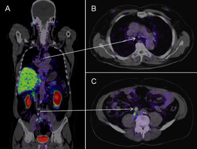
- Positron emission tomography (PET) scans, which use a radioactive substance to look for disease in the body. PET scans can show how body tissues are functioning.
- Biopsies, where doctors remove a sample of suspicious tissue to examine under a microscope. This is often the only definitive way to confirm whether cancer is present.
Each test has its own strengths and weaknesses. Doctors may order multiple different tests and compare the results to get a more complete picture before making a diagnosis. Generally CT scans are used as an initial screening test, while MRI, PET and biopsy results help confirm or rule out cancer.
When CT Scans are Recommended
Doctors generally recommend CT scans for cancer screening or diagnosis when there are signs or symptoms of cancer, or for patients who are at higher risk. According to the American Cancer Society, CT scans are commonly used to diagnose cancers of the lungs, liver, adrenal glands, kidneys, pancreas, prostate, bone, thyroid, and the brain (Gebai, 2023). They are often the best way to see tumors in the chest, abdomen, and pelvis. CT scans can show small tumors and be used to guide biopsy needles precisely into the abnormal area to obtain samples for analysis.
The U.S. Preventive Services Task Force recommends annual low-dose CT screening for lung cancer in adults aged 50–80 years who have a 20 pack-year smoking history and currently smoke or have quit within the past 15 years. This screening has been shown to reduce mortality from lung cancer (USPSTF, 2022). CT scans may also be recommended for patients with symptoms or higher risk factors for other cancers, such as family history.
However, most medical groups do not recommend routine CT screening for cancers in people without symptoms or higher risk. This is because false positives and incidental findings are common, which can lead to unnecessary anxiety and follow-up tests. Doctors will weigh the benefits and risks before recommending CT scans for cancer screening (Meadows, 2021).
Improving CT Scan Accuracy
There are several new developments that aim to improve the accuracy of CT scans for detecting cancer. One advancement is spectral CT, which uses multiple energy levels to provide better soft tissue contrast and characterization compared to conventional CT scans (1). This can improve tumor visibility in cancers like lymphomas and head and neck cancers (2).
Using intravenous contrast dyes can also enhance the visualization of tumors and vessels on CT scans. Certain dyes are taken up differently by normal tissue versus tumors, allowing radiologists to better see cancerous areas.
Radiomics is a new field that extracts quantitative imaging features from CT scans using data-mining algorithms. This big data approach helps reveal imaging biomarkers related to cancer genetics and prognosis. When combined with machine learning, radiomics can potentially improve diagnostic accuracy beyond visual assessment alone.
Artificial intelligence and deep learning software is also being developed to help radiologists evaluate CT scans faster and with higher accuracy. Studies have found AI can double evaluation speed and reduce errors in assessing scans for advanced cancers (3).
Conclusion
While CT scans have limitations, they can play an important role in cancer detection and diagnosis when used appropriately. CT scans may miss some cancers, especially smaller tumors or those obscured by dense tissue. However, they excel at detecting many cancers in the lungs, colon, liver, kidneys and other organs. When combined with a patient’s medical history and other diagnostic tests like PET scans or MRI, CT scans can help doctors determine if cancer is present and if it has spread. The accuracy of CT scans is improving with advances in technology.
Patients concerned about cancer should have an open conversation with their doctor about the appropriate screening and diagnostic tests based on their medical profile and risk factors. While CT scans can certainly detect many cancers, they may not be warranted in all cases and could lead to false positives. Doctors can determine if a CT scan, MRI, PET scan, biopsy or other test is most appropriate. With the right testing protocol tailored to the individual patient, doctors can often accurately diagnosis cancer and staging.