
CT (computed tomography) scans are medical imaging tests that use X-rays and computers to create detailed pictures of the inside of the body (https://www.cancer.gov/about-cancer/diagnosis-staging/ct-scans-fact-sheet). They are an important tool used by doctors to diagnose diseases and guide treatment. CT scans provide a fast and accurate view of the body and can detect cancers, strokes, injuries, and other health issues.
While CT scans provide many benefits, there are some concerns about potential cancer risks from radiation exposure (https://www.cdc.gov/nceh/radiation/ct_scans.html). This article will provide an overview of how CT scans work, the radiation dose from scans, evidence of cancer risks, factors that influence risks, weighing scan benefits vs risks, ways to reduce radiation exposure, scan alternatives, and key conclusions.
How CT Scans Work
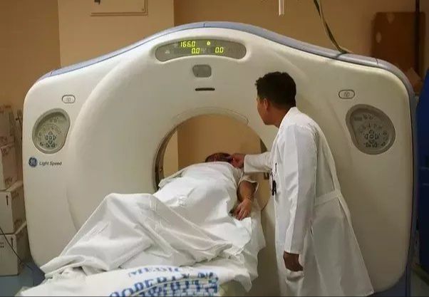
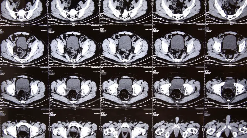
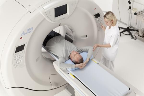
CT scans, also known as CAT scans, use a specialized X-ray machine to take multiple images or pictures of the inside of the body from different angles. The images are then combined by computer software to create cross-sectional views or “slices” of the area being scanned (Webmd).
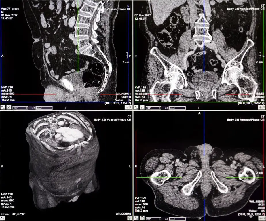
Instead of taking one X-ray image like a regular X-ray machine, a CT scan takes multiple images as it rotates around the body. This allows it to obtain more detailed information and construct 3D images of internal organs and other structures (Avala).
CT scans may use a contrast dye, which is either swallowed or injected into the bloodstream. The contrast dye helps highlight specific areas inside the body and improves the visibility of certain tissues and blood vessels (Webmd).
The X-rays used in CT scans involve higher amounts of radiation compared to regular X-rays. The rotation around the body allows the CT machine to capture a large series of narrow X-ray beams through the area being examined (Avala).
CT Scan Radiation Exposure
CT scans involve much higher radiation exposure compared to regular X-rays. According to one study, the average radiation dose from a CT scan is equivalent to about 100-500 chest X-rays. A chest CT scan emits about 100 times more radiation than a simple chest X-ray.
To put this in perspective, natural background radiation exposure is about 3 mSv per year. A chest X-ray is about 0.1 mSv, while a chest CT scan exposes you to about 7 mSv of radiation. A full body CT scan can be 10 mSv or higher.
So clearly, CT scans involve a much more intense radiation exposure than basic X-rays. However, it’s still generally less exposure than we get from natural background radiation over 1-2 years. The increased exposure is considered acceptable for diagnostic purposes when the benefits outweigh the small cancer risk.
Radiation and Cancer Risk
Ionizing radiation has enough energy to damage DNA inside cells by causing breaks in the DNA strands or interfering with DNA replication (https://books.google.com/books?id=3Fn-cyCymHoC&pg=RA1-PA614&lpg=RA1-PA614&dq=%22radiation+cancer+mechanism%22&source=bl&ots=Yum1SjpBrg&sig=ACfU3U0e-BsZ65VeeBj1xTmnlItzJJqvlQ&hl=en&sa=X&ved=2ahUKEwjhg97io7GDAxUl7rsIHboPBTAQ6AF6BAgJEAM). This can disrupt the cell’s ability to properly repair and replicate its DNA before dividing. If the damaged DNA is not properly repaired, it can lead to mutations or abnormalities in the new cells. Over time, accumulation of DNA damage and mutations can cause cells to grow out of control and develop into cancer.
The level of radiation exposure affects how likely this damage is to occur. Higher radiation doses are more likely to cause DNA breaks and mutations. Even low doses of radiation may slightly increase cancer risk by causing subtle DNA damage.
However, it’s important to note that the development of cancer is a complex, multi-step process. Radiation exposure increases cancer risk but does not automatically mean cancer will develop. The risk rises with higher radiation doses.
Evidence of CT Scan Cancer Risk
Several major studies have examined the potential cancer risk from CT scans. In a 2009 study published in the Archives of Internal Medicine, researchers estimated that 29,000 future cancers could be related to CT scans performed in the U.S. in 2007. The risk was found to be higher for younger patients.
Another major 2013 study in the British Medical Journal examined over 180,000 young people who underwent CT scans between 1985 and 2002 in the UK. The researchers found a positive association between radiation exposure from CT scans and increased risk of leukemia and brain tumors.
The authors of both studies concluded that CT scans should only be performed when medically justified, since they do carry a small but measurable risk of cancer. However, they emphasized that the benefits of diagnosing conditions correctly often outweigh these potential risks.
Risk Depends on Factors
The cancer risk from CT scans depends on several factors, including age, number of scans over time, and type of scan.1 Children have a higher lifetime cancer risk from radiation exposure compared to adults because their organs are still developing and they have many years of life ahead.2 Having multiple CT scans over time also increases cumulative radiation exposure and associated cancer risks.3
Not all CT scans deliver equal doses of radiation. CT scans of the abdomen and pelvis result in higher organ radiation doses than scans of the head or chest.1 Newer CT scanners also use lower dose protocols that reduce radiation exposure. Understanding these variables can help patients and doctors make informed decisions about CT scan risks and benefits.
Weighing Benefits vs Risks
When considering whether to get a CT scan, it is important to weigh the potential benefits against the potential risks. The benefits of a CT scan include detecting or ruling out serious medical conditions, guiding medical treatment, and providing peace of mind. However, there is a small potential risk of developing cancer later in life due to the radiation exposure from the scan.
It is best to carefully discuss the need for a CT scan with your doctor. Many medical organizations advise that CT scans be limited to cases where there is a clear medical benefit that outweighs the radiation risks. For many acute medical issues, especially in emergency situations, the benefits often do outweigh the small cancer risks from the radiation exposure. However, the risks may outweigh the benefits for excessive CT scans or when other good testing alternatives are available.
Doctors can help patients determine if a CT scan is truly necessary or if other tests like ultrasound or MRI could provide the needed information without ionizing radiation. It is also important to talk to your doctor about whether follow-up or repeat CT scans are required, as the radiation risks are cumulative. By carefully considering whether the CT scan is justified and necessary, patients and doctors can maximize the benefits while minimizing any potential long-term risks.
Reducing CT Scan Radiation
There are several ways to reduce radiation exposure from CT scans while still producing high quality images for diagnosis. Some strategies focus on advances in CT scanner technology and imaging techniques. Newer CT scanners can produce clearer scans with less radiation exposure. For example, one technique uses statistical modeling to reduce noise in low-dose scans (https://www.technologyreview.com/2010/08/02/201753/clear-ct-scans-with-less-radiation/). Size-based dose reductions aim to tailor the radiation dose based on the size of the patient, since less radiation is needed for smaller patients.
Other methods involve added shielding and protection. Bismuth shields can be placed over sensitive organs not being directly scanned to limit exposure. Lead shields also protect the patient by blocking scattered radiation. Proper shielding placement is important to avoid interfering with the scan. Some facilities provide lead-lined gowns to patients for extra protection.
By combining technological advances, size-based dosing, and shielding techniques, radiologists can optimize CT protocols to use only the minimum radiation needed for each patient. Ongoing research also continues to improve image quality while reducing radiation exposure from these important diagnostic scans.
Alternatives to CT Scans
While CT scans are a useful diagnostic imaging tool, the radiation exposure has led physicians and patients to consider alternatives when appropriate. Two of the main alternatives are ultrasound and MRI.
Ultrasound uses high-frequency sound waves to produce images of internal body structures. It does not use ionizing radiation so there is no exposure risk. Ultrasound is often used for imaging of the abdomen, heart, veins, arteries, joints, and during pregnancy. However, ultrasound does have limitations in visualizing some areas of the body.1
MRI (magnetic resonance imaging) also does not use ionizing radiation. It utilizes strong magnetic fields and radio waves to generate very detailed images of organs and tissues. MRI is preferred over CT for brain abnormalities, cancers, torn ligaments, spinal issues, and evaluating joints. The downside is MRI scans take longer than CT scans and can be unsuitable for patients with medical implants.
When considering alternatives to CT scans, doctors and patients should weigh the benefits against any disadvantages or limitations. Ultrasound and MRI avoid radiation but may not provide the same quality imaging as a CT scan for some conditions. The choice depends on the medical need, area being scanned, and patient factors.
Conclusion
CT scans are a useful medical imaging technology that provide important diagnostic information. However, the radiation exposure from CT scans does carry a small but measurable increased lifetime risk of developing cancer. The exact level of risk depends on many factors like age, scan protocols, and your background radiation exposure.
While CT scan radiation is concerning, in many cases the benefits outweigh the potential risks. It’s important to have an informed discussion with your doctor about whether a CT scan is recommended for your situation, and how to optimize the scan protocol to reduce unnecessary radiation exposure. With the help of your physician, CT scans can be used judiciously and appropriately.
The increased cancer risk from a single or occasional CT scan is generally small, particularly in adulthood. This risk is best minimized by avoiding excessive scans and ensuring each scan is optimized and warranted. Work collaboratively with your doctor to make personalized decisions regarding CT scans. Moderate, rational use of this technology can provide life-saving information that improves health outcomes.
