
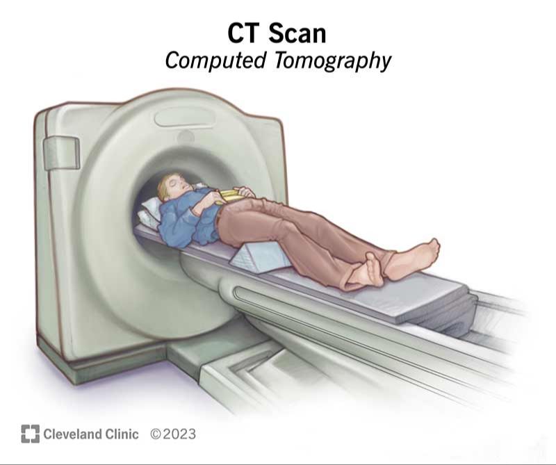
A CAT scan, also known as a CT (computed tomography) scan, is an imaging test that uses x-rays and computers to create detailed, cross-sectional images of the inside of the body. During a CAT scan, the patient lies on a table that moves through a large donut-shaped machine. X-rays are taken at multiple angles around the body and processed by the computer to generate images. Because it provides more detailed images of organs, bones, soft tissues and blood vessels than regular x-rays, CAT scans are often used to detect tumors and diagnose cancer.
What is a CAT Scan?
CAT scan stands for “computed axial tomography” scan. It is a type of radiography that uses X-rays to take cross-sectional images or “slices” of the body. A CAT scan can provide much more detailed images of soft tissues compared to a regular X-ray.
During a CAT scan, the patient lies on a table that slides into a large, circular opening of the CAT scan machine. The X-ray tube inside the machine rotates around the body and sends narrow beams of X-rays through the patient from many different angles. As the X-rays pass through tissues of different densities, the X-rays are absorbed or scattered at different rates. The X-ray detector on the opposite side of the body collects the X-ray data coming out of the body and sends it to the computer. The computer combines the data from the X-ray detector with the position of the X-ray tube to generate detailed cross-sectional images of the body.
The term CAT scan comes from the fact that the X-ray beam is rotated around a single axis of the body as the images are captured. This allows slice images of structures to be taken along that axis, called the axial or transverse plane. The computer stacks these axial slices together to recreate detailed 3D representations of structures inside the body for analysis.
How a CAT Scan Works
A CAT scan, also known as a CT or computed tomography scan, uses a combination of x-rays and computer technology to produce detailed images of the inside of the body. Here is a general overview of how a CAT scan works:
The scan is performed using a large donut-shaped machine called a CT scanner. The scanner contains an x-ray tube that rotates around the body and beams of x-rays are aimed at the area being examined. The patient lies on a table that slides into the center of the scanner.
As the x-rays pass through the body, they are absorbed in different amounts depending on the density of the tissue they go through. Denser tissues like bone absorb more x-rays than softer tissues. Detectors on the opposite side of the scanner measure the amount of x-rays that pass through the body.
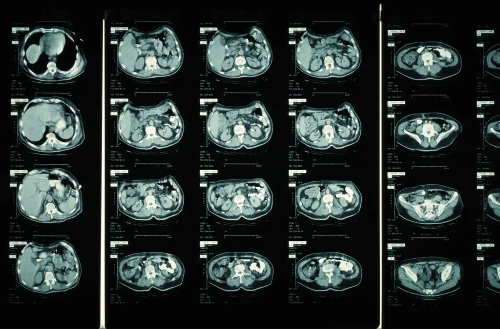
The scanner’s computer takes all these x-ray measurements and converts them into 2D cross-sectional images called slices. These image slices represent a thin section of the body. The computer then stacks the image slices together to generate a 3D image of the area.
So in summary, a CAT scan uses rotating x-rays and detectors to capture multiple image slices through the body. Advanced computer processing transforms this data into detailed 2D and 3D images.
What CAT Scans Can Detect
CAT scans, also known as CT or computed tomography scans, are often used to detect tumors and cancer in the body. The scans use X-rays and computers to produce cross-sectional images of the internal organs and tissues [1].
One of the main uses of CAT scans is detecting tumors, both benign and cancerous. The scans allow doctors to pinpoint the location of tumors and examine their size and shape. This helps determine the stage of cancers so doctors can plan the most effective treatments. CAT scans are considered one of the best imaging tests for detecting many types of cancers at an early stage when treatment is most successful.
In addition to tumors and cancer, CAT scans can effectively visualize other structures and conditions in the body. They excel at imaging the bones and detecting fractures or bone diseases like osteoporosis. CAT scans also provide excellent images of blood vessels, allowing doctors to check for blockages or aneurysms. They can also be used to evaluate organs like the liver, kidneys, spleen, pancreas and adrenal glands.
Overall, CAT scans are a versatile diagnostic tool. Their ability to create cross-sectional 3D images makes them invaluable for detecting a wide range of abnormalities and diseases in the body.
Using CAT Scans to Diagnose Cancer
CT scans can be an important tool for radiologists to analyze the body and identify potential tumors or cancerous growths (https://www.cancer.org/cancer/diagnosis-staging/tests/imaging-tests/ct-scan-for-cancer.html). The scans provide cross-sectional images that allow radiologists to examine both soft tissues and bones in detail.
When examining a CT scan, radiologists look for abnormal growths, changes in organ size or shape, inflamed lymph nodes, and other signs that may indicate cancer. Tumors and malignant growths often appear denser on CT scans compared to surrounding healthy tissue. Using contrast dye can make certain tumors stand out even more clearly.
However, CT scans do have some limitations in determining if a tumor is malignant. The scans themselves cannot confirm if abnormal tissue growth is cancerous – they can only identify areas of concern. A biopsy is needed to examine cells under a microscope and make a definitive cancer diagnosis.
Additionally, very small tumors or cancerous cells may not be detectable on a CT scan. The resolution limits of the scans mean some cancers can be missed, especially in early stages. So a “clear” CT scan does not necessarily mean there is no cancer present.
Overall, CT scans are an invaluable tool for locating potential tumors in the body and guiding doctors to areas that require further testing. But the scans cannot provide a conclusive cancer diagnosis on their own (https://www.cancercenter.com/diagnosing-cancer/diagnostic-imaging/ct-scans). A full workup with clinical correlation is needed to confirm malignancy.
Advantages of CAT Scans
CAT scans have several major advantages over other imaging techniques:
- They are non-invasive and do not require incisions or injections, unlike more invasive techniques like biopsies.
- CAT scans produce highly-detailed 3D images that provide more information than standard x-rays. The images allow doctors to view cross-sectional slices of the body (Mayo Clinic, 2022).
- Scan times are very quick, usually 5-10 minutes for one area of the body (RadiologyInfo.org, 2022). This allows doctors to scan multiple areas in one appointment.
- CAT scans can detect issues not visible with x-rays or MRI, like small tumors or organ damage.
Disadvantages of CAT Scans
While CAT scans provide detailed images of the inside of the body, they do have some downsides to consider. Here are some of the main disadvantages of CAT scans:
Expensive. CAT scans are significantly more expensive than regular X-rays. According to the U.S. National Library of Medicine, a chest X-ray costs less than $200, while a chest CT scan can cost over $1,000. The scans require large, sophisticated machines and specially trained technicians to operate them. Many insurance plans limit coverage of CAT scans or require prior authorization to control costs.
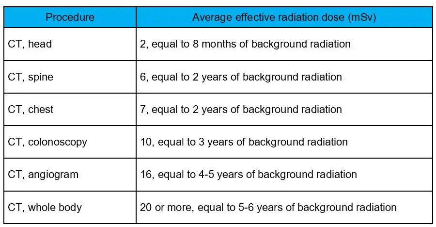
Radiation exposure. CAT scans use ionizing radiation in the form of X-rays to generate images. This radiation exposure may slightly increase a person’s overall lifetime risk of developing cancer, according to the U.S. Food and Drug Administration. However, the benefit of an accurate diagnosis usually outweighs this small risk.
Can miss small tumors. While CAT scans are excellent at detecting large abnormalities, their resolution limits their ability to see very small tumors or growths, according to WebMD. A 5 mm lesion may be undetectable on a scan. CAT scans may also have difficulty finding tumors located in fatty tissue. Additional tests like MRIs or biopsies may be necessary.
Risks from contrast dye. Many CAT scans use an intravenous contrast dye to enhance the visibility of internal structures and tissues. This dye can cause mild to severe allergic reactions in some people, reports the National Center for Biotechnology Information. Kidney damage is also a rare risk.
When a CAT Scan is Recommended
A CT scan may be recommended for a variety of reasons. Some common indications that a doctor may order a CT scan include:
- Cancer screening or staging, especially for cancers like lung, liver, and pancreatic cancers (https://www.mayoclinic.org/tests-procedures/ct-scan/about/pac-20393675)
- Evaluation of symptoms that could indicate cancer, like an unexplained lump or mass, unexplained weight loss, or bleeding (https://www.webmd.com/cancer/what-is-a-ct-scan)
- Monitoring cancer treatment and evaluating response to therapies (https://www.lompocvmc.com/blogs/2021/december/12-reasons-you-may-need-a-ct-scan/)
- Looking for spread or metastasis of known cancer (https://www.mayoclinic.org/tests-procedures/ct-scan/about/pac-20393675)
CT scans are often used to screen high-risk patients, like those with a family history of cancer, smokers, or those exposed to radiation or carcinogens. They may also be used to evaluate vague symptoms that could possibly indicate cancer in its early stages.
Preparing for a CAT Scan
Proper preparation before a CAT scan is important to ensure the scan goes smoothly. According to the University of Connecticut Health Center [1] and WakeMed [2], there are a few key steps to take:
You will likely be asked to avoid food for several hours before the scan, especially if contrast dye will be used. This helps prevent nausea and vomiting during the procedure. Follow your doctor’s specific instructions on fasting.
When you arrive for the scan, you will change into a hospital gown. You’ll need to remove any jewelry, eyeglasses, or metal objects that could interfere with the imaging. Inform staff beforehand if you have any medical implants containing metal.
If your scan requires an injected contrast dye, you may need to drink an oral solution beforehand. This highlights certain tissues and blood vessels for the scan. The taste and preparation varies depending on the type of dye used.
These steps allow for the clearest possible CAT scan images, leading to the most accurate diagnosis.
Conclusion
CAT scans play an important role in detecting tumors and diagnosing cancer due to their ability to create detailed cross-sectional images of the body. The key points covered in this article about using CAT scans for tumor detection include:
– CAT scans (also called CT scans) use a series of X-ray images and computer processing to create 3D images that can precisely show the size, shape and location of many types of tumors.
– CAT scans excel at detecting tumors in the chest, abdomen and pelvis, including cancers of the lung, liver, kidney, ovaries and pancreas. They can often detect tumors at earlier stages compared to other methods.
– The detailed images produced by CAT scans allow doctors to determine if a mass is likely cancerous, see how far cancer may have spread, guide biopsies, and monitor treatment effectiveness.
– While disadvantages like radiation exposure exist, a CAT scan’s benefits often outweigh these risks in cases of suspected cancer. Patients should discuss options with their doctor.
– With their detailed 3D views and ability to detect even small tumors, CAT scans have become a vital tool for diagnosing many types of cancer and detecting tumors throughout the body.
